Bats can carry various diseases, including many which are transferable to humans. A recent study published in the Journal of Animal Ecology investigated disease extent, seasonality, and mechanisms of transmission among Malagasy fruit bats. Lead author Dr Cara Brook (Princeton University and UC Berkeley) explains more about the paper.
Bats (order Chiroptera) have received much attention in recent years for their roles as reservoirs for some of the world’s most virulent zoonoses—including Ebola and Marburg filoviruses and Hendra and Nipah henipaviruses (Calisher, Childs, Field, Holmes, & Schountz, 2006) —which they appear to host without experiencing disease. Numerous empirical studies have demonstrated seasonality in bat viral and immunological dynamics, which coincide with the annual chiropteran birth pulse and may underpin observed seasonality in cross-species emergence of bat-borne zoonoses (Amman et al., 2012; Baker et al., 2014; Plowright et al., 2015, 2008; Schmidt et al., 2017). Previous theoretical work has attempted to recapture these seasonal dynamics using models encoding a suite of diverse transmission mechanisms, but application of these models to longitudinal field data has, to date, been rare. In a recent paper published in the Journal of Animal Ecology, we coupled field and modeling approaches to investigate the question of bat henipa- and filovirus seasonality on the Eighth Continent island nation of Madagascar.

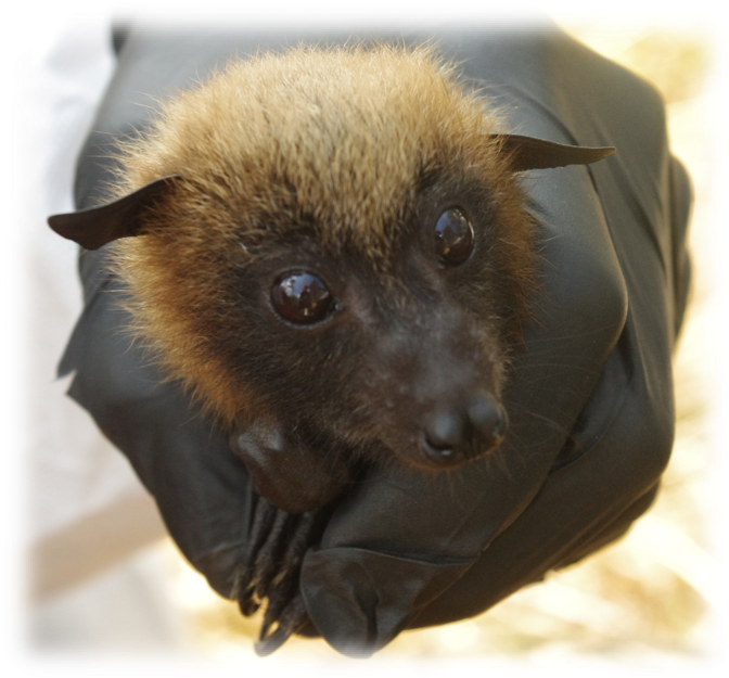
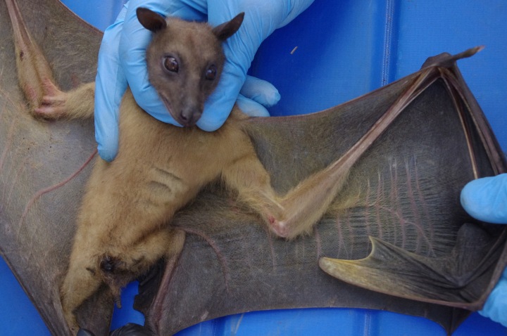
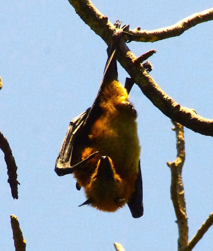
During the course of my PhD and in close collaboration with Christian Ranaivoson, a Malagasy doctoral student at the University of Antananarivo and the Institute Pasteur of Madagascar (and the second-author on this paper), we field-captured and serum-sampled over 800 fruit bats from three endemic Malagasy fruit bat species (Pteropus rufus, Eidolon dupreanum, and Rousettus madagascariensis) across all months of the year. From a subset of larger-bodied P. rufus and E. dupreanum, we extracted teeth under anesthesia, which were sliced and stained, with layers counted to determine each bat’s age. Serum underwent Luminex-based antibody assay against known filo- and henipavirus antigens.
Deciphering serology
Antibodies are memory proteins which the mammalian immune system uses to neutralize harmful viruses entering the body. Assays such as our Luminex measure the extent to which antibodies in serum bind the antigens against which they are challenged. The presence of antibodies specific to an antigen thus provides evidence of past exposure of the host from which the sample is taken to the viral antigen against which the sample is challenged.

Our study reports the first evidence of Ebola “seropositivity” (antibody positivity) in any wild animal in Madagascar —specifically, in P. rufus and R. madagascariensis—in addition to the first evidence of Cedar virus (a bat-exclusive henipavirus) seropositivity in Madagascar—in E. dupreanum and R. madagascariensis. We further confirm previous records of seropositivity to Hendra and Nipah virus antigens in E. dupreanum and P. rufus (Iehlé et al., 2007) and additionally report the first evidence of Hendra and Nipah virus seropositivity in R. madagascariensis.
Because we lack specific antigens for viruses of Madagascar fruit bats, seropositivity was determined based on binding of Malagasy bat serum antibodies to antigens from African Ebola and Asian and Australian Nipah and Hendra viruses. As antibodies can sometimes bind antigens from similar viruses, it is possible that our seropositive fruit bats are, in actuality, hosting unique Ebola-, Hendra-, and/or Nipah-related viruses that have not yet been described. We are working actively to identify and isolate these viral genotypes to date.
Consistent with previous reports in the literature, we witnessed seasonal changes in population-level seroprevalence (the proportion of bats testing antibody positive) to Nipah virus in E. dupreanum and Ebola virus in P. rufus. Seroprevalence peaked at the height of the fruiting season and at the gestation/lactation transition for bats in our system. Data from individual E. dupreanum that were captured twice across our study supported population-level trends: female serotiters increased across gestation and declined post-lactation, while male serotiters increased throughout the fruiting season, then decreased across the dry season. Seasonal trends in body mass similarly mapped on to the reproductive calendar for females and the nutritional calendar for males.
Elucidating transmission
Traditionally, dynamical insights in disease ecology and public health have been largely derived via fitting of compartmental transmission models in the Susceptible-Infectious-Recovered (SIR) framework to times series of infectious cases. Under such a framework, hosts are classed into infection state categories (S-I-R), and parameters corresponding to transmission or recovery rates are optimized to produce the best recapitulation of the data. In the case of wildlife infections, where time series case data are difficult to obtain, we use tooth-derived age information, paired with serology (corresponding to the “recovered”, “R”-class) to improve modeling inference.

For both Nipah virus in E. dupreanum and Ebola virus in P. rufus, seroprevalence was high in neonates, low in juveniles, increased with age across early adult years and finally waned again in older bats. Using a matrix-based approach, which enabled us to track bat ages within each infection state in our system, we were best able to recapitulate these data with an MSIRN model structure, by which bats passed from a state of maternally immune (M) to susceptible (S) to infectious (I) to recovered (R) before finally settling in to an “N”-class, representing “non-antibody-mediated immunity.” We modeled bats in this age class as antibody-negative via our Luminex but nonetheless immune to reinfection via some other immune-protective mechanism, consistent with some previously-reported experimental infections in the literature.
Our work greatly advances the conversation in bat virus disease ecology, but there remain many unanswered questions in this system and this field. For example, our MSIRN model assumes that the older bats become uniformly seronegative but nonetheless immunized for life—but we know from seasonal monitoring of serology that serotiters are dynamic for individual bats. It is possible then that older bats may go through seasonal cycles of seropositivity and/or actively shed virus in excretia, a process not explicitly modeled here. In the future, more finescale longitudinal sampling of recaptured individuals will facilitate construction of models incorporating within-host processes. This article represents the culmination of much of the work emerging from my PhD—but nonetheless launches me into a lifetime of questions and discoveries ahead.

More Info:
Amman et al. (2012). Seasonal pulses of Marburg virus circulation in juvenile Rousettus aegyptiacus bats coincide with periods of increased risk of human infection. PLoS Pathogens, 8(10), e1002877. doi:10.1371/journal.ppat.1002877
Baker et al. (2014). Viral antibody dynamics in a chiropteran host. Journal of Animal Ecology, 83(2), 415–428. doi:10.1111/1365-2656.12153
Brook et al. (2019) Disentangling serology to elucidate henipa‐ and filovirus transmission in Madagascar fruit bats. Journal of Animal Ecology. doi:10.1111/1365-2656.12985
Calisher et al. (2006). Bats: Important reservoir hosts of emerging viruses. Clinical Microbiology Reviews, 19(3), 531–45. doi:10.1128/CMR.00017-06
Iehlé et al. (2007). Henipavirus and Tioman virus antibodies in Pteropodid bats, Madagascar. Emerging Infectious Diseases, 13(1), 159–161.
Plowright et al. (2015). Ecological dynamics of emerging bat virus spillover. Proceedings of the Royal Society B, 282(1798). doi:10.1098/rspb.2014.2124
Plowright et al. (2008). Reproduction and nutritional stress are risk factors for Hendra virus infection in little red flying foxes (Pteropus scapulatus). Proceedings of the Royal Society B: Biological Sciences, 275(1636), 861–9. doi:10.1098/rspb.2007.1260
Schmidt et al. (2017). Spatiotemporal fluctuations and triggers of Ebola virus spillover. Emerging Infectious Diseases, 23(3), 415–422. doi:10.3201/eid2303.160101
